B. Nielsen L, B. Svensson R, U. Fredskild N, et al.
Scand J Med Sci Sports. 2023;33:2585-2597
Background
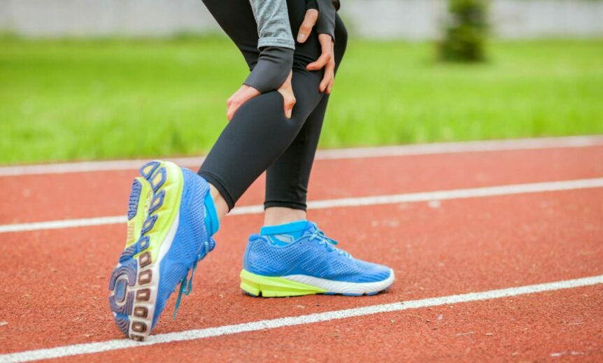
Muscle strain injuries in the human calf muscles are frequent sports injuries with high recurrence. Potential structural and functional changes in the medial head of the musculus gastrocnemius (GM) and the associated aponeurosis are not well documented.
Purpose
To test whether a GM muscle strain injury affects muscle fascicle length, pennation angle, and the morphology of the deep aponeurosis at rest and during muscle contraction long time after the injury. Additionally, electromyography (EMG) of the GM and the soleus muscle during a unilateral heel rise was measured in the injured and uninjured calf.
Methods
GM fascicle length, pennation angle, and aponeurosis thickness was analyzed on dynamic ultrasonography (US) recordings in 10 participants with a chronic calf strain. In addition, US images taken across the distal portion and mid-belly of the GM were analyzed at three different ankle positions. EMG recordings were obtained during a unilateral heel rise.
Results
The pennation angle of the injured distal GM was significantly larger compared to the uninjured GM in the contracted, but not the relaxed state. Pennation angle increased more in the injured compared to the uninjured GM during contraction. Fascicle length was shorter in the most distal portion of the injured GM. Fascicles at the distal portion of the injured GM showed a pronounced curvilinear shape as the muscle contracted and the aponeurosis was enlarged in the injured compared to the uninjured GM. The ratio between GM and soleus EMG activity showed a significantly higher relative soleus activity in the injured compared to the healthy calf.
Conclusion
The greater change in pennation angle and curvilinear fascicle shape during contraction suggest that a long-term consequence after a muscle strain injury is that some muscle fibers at the distal GM are not actively engaged. The significantly enlarged aponeurosis indicates a substantial and long-lasting connective tissue involvement following strain injuries.